Dr. Naoki Kogo and Prof. Nael Nadif Kasri
Department of Human Genetics/Cognitive Neuroscience, Radboud University Medical Centre, Nijmegen, Netherlands
Background
Dr. Naoki Kogo is a visiting researcher in the lab of Prof. Nael Kasri, participating in projects to study the neural properties of network dysfunction in neurodevelopmental disorders. This lab uses human induced pluripotent stem cells (iPSCs) to form neural networks, and then applies electrophysiological and optical techniques to study the function of these networks and organoids, using calcium imaging and voltage imaging.
These models are highly relevant to medical research as they are based on human cells, and can help to elucidate the mechanisms behind neurological disease and how dysfunction in neural networks can occur. Both calcium imaging across large neuronal populations and voltage imaging to see individual action potentials are used to get a complete view of functionality.
Challenge
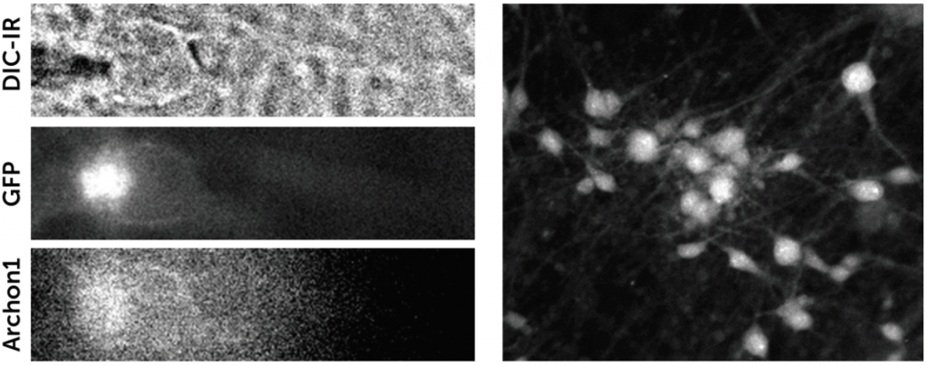
Combining both calcium and voltage imaging means this research requires a flexible yet powerful detector. Calcium imaging requires a large field of view (FOV) at a high resolution in order to see waves of activity across large neuronal populations at low magnification (x20), and voltage imaging requires high speed and high sensitivity imaging to observe fast dynamic events within smaller numbers of neurons, at high magnification (x60).
Dr. Kogo mentioned the lab had used an older model of camera that could not perform the voltage imaging, so a new detector was required.
We are happy with the performance of the [Prime BSI Express], we are now able to capture voltage signals in our neural networks.
Solution
The Prime BSI Express is a highly flexible camera that is well suited to both calcium and voltage imaging. This CMOS has a large FOV and a balanced 6.5 μm pixel, well suited to calcium imaging across large numbers of neurons, with the CMS mode providing an excellent signal-to-noise ratio.
In addition, the high 95 fps and low read noise of the Prime BSI Express allow it to perform voltage imaging, operating at a high speed with high sensitivity. Through the use of regions of interest and binning, the Prime BSI Express can maximize signal collection while minimizing noise, collecting even weak fluorescent signals at high speed.