Dr. Sonia Leonardelli, Prof. Michael Hölzel
Hölzel Lab, Institute of Experimental Oncology, University Hospital Bonn
Background
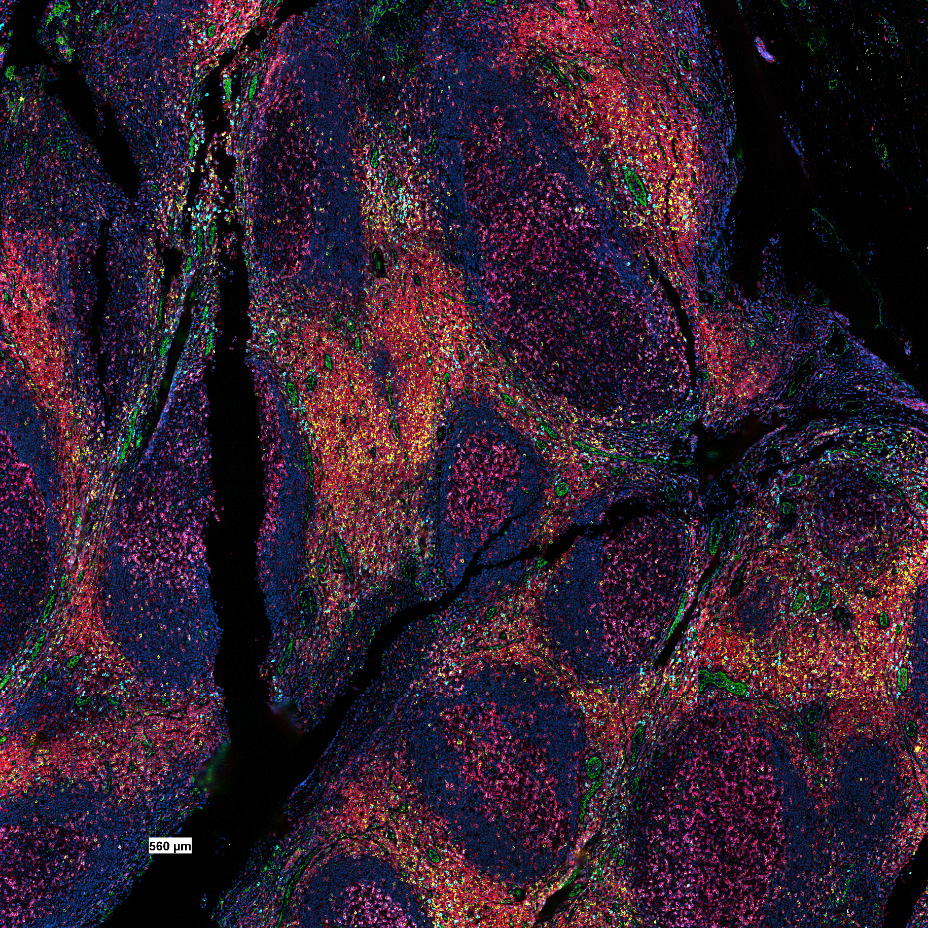
Dr Sonia Leonardelli of the Hölzel Lab performs fluorescent imaging of a range of different tumour tissue samples, one of the main projects being to study cell-cell interactions in adenocarcinoma.
The fluorescence imaging in this lab using the CODEX® system from Akoya Biosciences® with a Zeiss microscope in order to take multiple fluorescence images with multiple different fluorophores, which can then be collated and output with all the fluorophores in the same image, up to 60 different types. Images from these experiments typically have up to 8 different fluorescent markers and are dense, high content images that reveal details about the tumour microenvironment.
Imaging tumour samples with these multiplex fluorescence methods allows Dr Leonardelli to see what interactions are occurring, how the tissues respond to drugs, and how tumour malignancy can develop. This will further allow for the development of biomarkers to see how tumours develop and how they respond to different therapies.
Challenge
The CODEX® system has a 10x or 20x objective, meaning that while images can be acquired across a large field of view, they are limited on their resolution due to the lower objective numerical aperture. A high-resolution camera with smaller pixels will be needed in order to acquire details on the cellular level in order to see cell-cell interactions.
Due to the wide range of fluorophores used (including DAPI, Cy3, Cy5, and Cy7) any detector used with this system would need a high quantum efficiency across a wide range of wavelengths, from blue to near-infrared, in order to use the most fluorophores without spectral overlap. A high sensitivity camera would also allow for a reduction in illumination intensity to avoid bleaching.
We chose [the Prime BSI] due to the high resolution and high sensitivity, especially in the infrared as the Cy7 gives very good images, we are very happy.
Solution
The Prime BSI is a great solution for this multiplex fluorescence imaging, as it offers both high sensitivity and high resolution. Dr Leonardelli stated that she was able to see cell-cell interactions due to the high resolution of the Prime BSI, across the multiple fluorophores available. The Prime BSI has 95% peak quantum efficiency, and high sensitivity across a wide range of wavelengths from UV to near-infrared, in order to acquire data from the various fluorophores in these multiplex experiments.
In addition, with CODEX® software optimization, Dr Leonardelli will also be able to reduce the illumination intensity and make use of the high sensitivity of the Prime BSI.