Dr Brian Patton, Mr Petros Hadjichristodoulou
Department of Physics, University of Strathclyde, Glasgow
Background
Dr. Brian Patton and Ph.D. candidate Petros Hadjichristodoulou are involved in microscopy development and incorporating technologies into imaging. One such technology is the use of nanodiamonds (NDs), these contain natural defects known as nitrogen-vacancy centers that can be modulated by illuminating the NDs, which will emit fluorescence. NDs are also non-toxic and photostable, and as such are used by the lab of Dr. Patton as biological probes for thermometry, measuring the temperature at a cellular level, even measuring temperature gradients across a whole cell population.
Dr. Patton explains his research, “Our first ND application will be thermometry, cells generate heat as a by-product of chemical processes, and so cellular activity can be measured via thermometry. This is a huge step towards more challenging measurements, such as measuring magnetic fields generated by biological activity, so once thermometry measurements are feasible you can look at sensitivity, and get interesting answers to biological questions.”
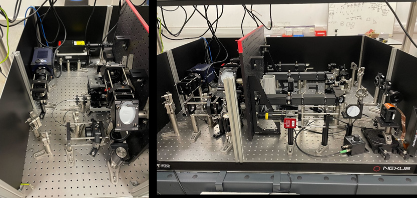
One current project for Dr. Patton is the design of a novel technique that combines several factors, namely using ND thermometry to study entire nematode worms (C. elegans) at the nanoscale using light-sheet microscopy with adaptive optics.
Challenge
While C. elegans have been studied with ND thermometry in widefield, or imaged using light-sheet microscopy, and light-sheet systems have been developed that use adaptive optics, the combination of light-sheet imaging, adaptive optics, and optical thermometry has not been done before. This results in a highly complex and demanding technique that presents unique and potentially unforeseen challenges.
When using a light-sheet, typical challenges involve the need for high resolution across a large field of view, while maintaining a low noise level for high sensitivity. Alongside this, when imaging large volumes it is important to have a high-speed detector in order to capture a sufficient number of volumes per second, especially with whole-organism light sheet.
The speed and QE of the [Prime BSI] were really good, as well as the fact that it extended into the NIR emission wavelength range for the anodiamond.
Solution
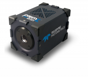
An ideal camera solution is the Prime BSI sCMOS, which features high resolution (2k x 2k sensor with 6.5 μm pixel), a large FOV, and fast imaging speeds. The Prime BSI is a high sensitivity sCMOS, featuring both high quantum efficiency (QE) for maximizing signal collection across a wide range of wavelengths, and low-noise CMS mode for minimizing noise levels.
Dr. Patton shared his thoughts on the Prime BSI, “The final version of our system on the table was designed for the Photometrics [Prime BSI]… the speed and QE we thought were really good, as well as the fact that it extended into the emission wavelength range for the ND.”
“Our volumetric imaging requires speed, so a CMOS is a natural choice. The Prime BSI matches particularly well with our required performance. In particular, the dedicated PCIe board allows us to stream the data from large datasets directly to storage, and this allows us to do the post-processing. This is helpful when developing and debugging new imaging approaches and when imaging moving animals. For future development, the range of acquisition modes will allow us to try algorithmic improvements, and the sensitivity increases the chances of success when in low signal-to-noise regimes.”
Mr. Hadjichristodoulou also told us about software, “We are able to run the [Prime BSI] using Python, and downloaded the Python wrapper from the Photometrics website. The PVCAMtest software is also great as you can access all the functionality of the camera as well before implementing it in software.”