Dr. Rodrigo de Campos Perin
Laboratory of Neural Microcircuitry (LNMC), École polytechnique fédérale de Lausanne, Switzerland
Background
The EPFL houses the Laboratory of Neural Microcircuitry (LNMC), which is dedicated to researching and unraveling the structure and function of neural microcircuits, particularly in the neocortex. This group is headed by Prof. Henry Markram and includes research scientist Dr. Rodrigo de Campos Perin, who is working with a combination of fluorescence microscopy and patch clamping for intracellular electrophysiological recordings. This work often uses multi-patch electrophysiology in order to record from multiple neurons.
Challenge
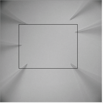
When performing patch clamping – especially multi-patching – with live neurons it is vital to have a good view of the field in order to accurately position the multiple electrodes in relation to cells. Dr. Perin noted that: “Keeping four electrodes in the field of view with our previous camera proved challenging in our usual experiments”. This combination of fluorescence and brightfield to position electrodes means that cameras need a high dynamic range to display both high and low signals without saturation, and a high resolution is needed to more accurately determine which cellular features are being patched and recorded.
“We recently started using the [Prime] BSI camera as a solution that allowed us to accelerate and make experiments more reliable in multiple ways… greatly improving the reliability in identifying and recording from fluorescently-labeled cells.”
Solution
Dr. Perin is now using the Prime BSI sCMOS camera for this application, and benefiting from the large field of view and excellent resolution. As mentioned by Dr. Perin: “We recently started using the BSI camera as a solution that allowed us to accelerate and make experiments more reliable in multiple ways.” The BSI enabled imaging of fluorescent signals while also positioning patch clamp electrodes, improving the reliability in identifying and then recording fluorescently-labeled cells.
In addition, the 11- and 16-bit modes of the BSI offer a high dynamic range, which Dr. Perin said: “Helps us concentrate on experiments without needing to adjust light intensity several times when performing electrophysiological recordings. It also provides the added advantage of more clearly visualizing anatomical features without the image saturation we experienced with our previous camera.”
The large field of view of the BSI now allows for six or more electrodes to be accommodated at high magnification, and makes it easier to target cells of interest even if they are deeper in the tissue, requiring fewer position adjustments and making experiments faster. Finally, the BSI’s 6.5 µm pixel offers excellent resolution, meaning that regions of interest can be expanded without loss of image quality, an approach much preferred to changing objectives or optically zooming as this can mechanically interfere with recordings.