Dr. Kyle Douglass, Post-Doctoral Researcher
The Laboratory of Experimental Biophysics EPFL
Suliana Manley Lab, Lausanne, Switzerland
Background
Dr. Kyle Douglass, a research scientist at the EPFL, has spent the past several years developing high-throughput and automation methods for super-resolution fluorescence microscopy. The Laboratory of Experimental Biophysics, which is led by Prof. Suliana Manley, uses these techniques to study the structural biology of multi-protein complexes such as chromatin foci, the bacterial division machinery, and the centrosome.
From the perspective of the technology, these structures share a common theme in that they require large datasets of high quality images to computationally combine into a structural model which can possibly consist of one or more disordered components. It is therefore imperative to acquire as much data as possible and to ensure that it meets the exacting standards required by the computational reconstruction pipelines.
Challenge
These multiprotein complexes are well-suited to super-resolution approaches like STORM and PAINT because they are too small to see with traditional light microscopy. Furthermore, their rich and often heterogeneous composition precludes a complete study with electron microscopy , but this problem is easily overcome with multi-color super-resolution. Unfortunately, not every available dye is bright and stable. The quality of the measurements depends critically on how many photons can be recorded from each dye molecule, which means that the protein maps that are reconstructed from weak dyes will suffer from a loss of resolution and quality.
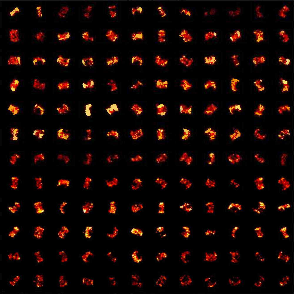
We can now more precisely locate each fluorescent dye that is targeting a protein within a complex [with the Prime 95B Scientific CMOS camera]. This has the effect of improving the resolution of our structural models, allowing us to see details inside these complexes that we could not before.
Solution
Because every photon counts, Douglass and colleagues upgraded their cameras to the Photometrics Prime 95B. The high sensitivity and large field of view allows the researchers to simultaneously image numerous structures at the same time while capturing even more photons than before. The increased throughput and quality of the data is paying dividends.