Mr Emil Kind and Prof. Mathias Wernet
Neurobiology and Neural Circuits, Institute of Biology, Freie University, Berlin
Background
Emil Kind is a PhD student in the lab of Prof. Wernet, which focuses on neural circuitry, especially circuits involved in navigation and orientation behaviour. Their studies span from looking at neuroanatomy on a cellular level to behaviour on the organism level using the fruit fly Drosophila as a model system.
There are special regions within the eye of the fruit fly used for orientation by looking at the sky. The lab of Prof. Wernet studies the downstream circuits in the fly brain, identifying and studying cells to see how they influence behavior.
Besides imaging the brain using confocal and multiphoton systems, Prof. Wernet’s lab also performs functional calcium and/or voltage imaging on ex vivo brain cultures. There are also further plans to move into optogenetics and use electrical stimuli.

Challenge
Mr Kind wants to use GCamp indicators to visualize activation and inhibition in neural brain cultures, but this can be very difficult and a high sensitivity camera is a necessity, especially when the experiments benefit from low-intensity illumination.
High-speed imaging is desirable in order to understand neural function, and high-resolution imaging allows for a better description of neural circuitry. The range of experiments requires solutions that are both flexible and powerful.
The base signal I am receiving is very nice, the pixel size and quantum efficiency of the [BSI Express] is really good.
Solution
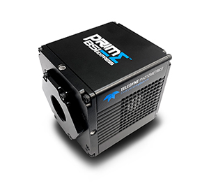
The Prime BSI Express is a high-speed, high-sensitivity solution for calcium imaging. Imaging at 95 frames a second across the full sensor, the BSI Express can capture neuronal function with ease. As Mr Kind says: “The [BSI Express] is just perfect, I don’t have any issues with the camera. I use a 60x objective and I can see all my neuronal population within the field of view.”
The 6.5 μm pixel of the BSI Express allows for flexible experimentation and optimized Nyquist sampling at 60x magnification, allowing for great spatial resolution when looking at individual neurons and whole slices containing dense circuitry.

