
Neuroscience and Electrophysiology
Electrophysiology is a method that allows us to study the function of electrically active cells, such as neurons. Research on the physiology of neurons and the nervous system is often accomplished at the cellular level using microscopy techniques or further techniques such as patch clamping.
Advanced imaging methods in neuroscience permit us to image deep into brain tissue with high spatial and temporal resolution. It is even possible, using advanced optogenetic methods, to optically interrogate cells to discover more about their function.
Cameras for neuroscience applications can have very different specification requirements based on what is being investigated. Cellular activity as a response to electrical stimulation or changes to membrane potentials, for example, can happen extremely quickly, so high frame rates can be of high importance. Many dyes used in electrophysiology are difficult to visualize because of a low quantum efficiency at the fluorophores emission wavelength. Therefore, a camera with a high quantum efficiency over the entire visible wavelength range may also be desired.
Finally, electrophysiology applications often require minimal electronic noise and vibrations to acquire high-quality data so the electrophysiologist requires tight control of camera noise.

Retiga ELECTRO
The Retiga ELECTRO is specifically designed for electrophysiology.
The high broadband quantum efficiency delivers the sensitivity required for high contrast imaging of cells and tissues, making the manipulation of micropipettes in their contact with cell membranes and tissues significantly easier to perform.
Fanless temperature regulation using a thermoelectric cooling system cools the sensor to 0oC without adding additional vibration to the system. This means that electrophysiology experiments no longer need to compensate for this additional noise.
Incorporated grounding pins allow the camera to be grounded within the rig to reduce the buildup of static electricity in electrical systems.
Finally, in-built Intelligent Quantification and Defective Pixel Correction technologies improve signal to noise for long exposures.

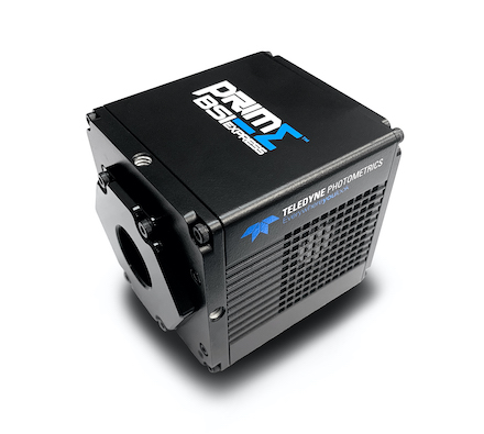
Prime BSI Express
High sensitivity, 95% quantum efficient, sCMOS camera with 6.5 µm pixels and 1.0 e– read noise.
The Prime BSI Express delivers the perfect balance between high-resolution imaging, sensitivity and speed. Nyquist spatial sampling is achieved at 60x magnification with no optical correction and the 45,000 e- linear full well capacity is 50% higher than other sCMOS cameras for the greatest dynamic range.
Read noise is minimized to just 1.0 e-, ideal for visualizing the smallest change in signal intensity, and with sCMOS speed the fastest events can be captured.
When the ELECTRO isn’t enough, the Prime BSI Express provides the necessary boost in sensitivity and speed.


Kinetix
High sensitivity, 95% quantum efficient sCMOS camera with an incredibly high 400 fps full-frame speed and a massive 29.4 mm diagonal field of view.
When speed is of vital importance, the Kinetix significantly outperforms typical sCMOS devices. With a full-frame framerate of 400 fps and a 10 megapixel sensor, the Kinetix delivers over 4000 megapixels/second.
The high quantum efficiency and low read noise combined with the balanced 6.5 µm pixel size also delivers the sensitivity needed to get the highest image quality from your electrophysiology system.

Customer Stories
NIR/VIS Brain Imaging
“I´d like to buy one more, please!”
High-Speed Voltage Imaging
“The Kinetix is the perfect solution for our requirements to detect voltage signals in vivo because it is the optimal solution – namely being fast and sensitive.”
Multi-Clamp Electrophysiology
“Very happy with [the Prime 95B], I was having problems before and this was a great solution, and probably the best optics I have.”